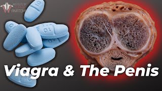
Published On Premiered Feb 14, 2023
The penis is a male external genital organ which is part of both the urinary and reproductive systems. The penis can be divided into three parts – the root, body and glans.
The root is not visible externally. It is the fixed part of the penis, located in the superficial perineal pouch of the pelvic floor. The root contains two crura and the bulb of the penis, which are erectile tissues, as well as the ischiocavernosus and bulbospongiosus muscles. Erectile tissues are tissues that engorge with blood during sexual arousal to create an erection. The two bulbospongiosum muscles are associated with the bulb of the penis and contract to empty the spongy urethra of residual urine or semen. It also contracts during orgasm and ejaculation. The two ischiocavernosus muscles surround the crura and contract to force blood distally from the crura’s cavernous spaces into the corpora cavernosa to add rigidity to an erection.
The body of the penis is suspended from the pubic symphysis, which is a joint between your right and left pelvic bones. It is composed of three columns of erectile tissue. Extending from the two crura in the root of the penis are a pair of corpora cavernosa, found along the sides of the penis. Extending from the bulb in the root of the penis is the corpus spongiosum, which runs along the bottom of the penis. The corpus spongiosum contains the urethra, which is the tube which allows for exit of urine or semen. Note that the corpus spongiosum fills with blood to a lesser extent than the corpora cavernosa during an erection so that the pressure does not result in the urethra becoming occluded. This way, semen can make their way through during ejaculation.
Finally, the glans is the penis’ most distal portion. It is a distal expansion of the corpus spongiosum and features the external urethral orifice, which is the opening of the urethra.
Erectile tissue has fascial coverings. The dartos fascia are found in the penis and scrotum, and the portion in the penis is called the superficial fascia of the penis. The part of the fascia in the scrotum is called the dartos proper. Note that the dartos fascia is continuous with Colles fascia in the perineum and Scarpa’s fascia in the abdomen. Going deeper, we see Buck’s fascia, also known as the deep fascia of the penis. It is a continuation of the deep perineal fascia, and holds the three erectile tissues together. Finally, beneath the deep fascia is the tunica albuginea, a strong fascia surrounding each cavernous body that is fused at the midline of the penis. Loose connective tissue connects the fascias to the overlying skin.
Two ligaments support the root of the penis – the suspensory ligament, which connects to the pubic symphysis, and the fundiform ligament, which runs down the linea alba, around the penis, and then attaches to the pubic symphysis.
The skin of the penis is more heavily pigmented than skin of the rest of the body. The foreskin, or prepuce, is a double layer of skin and fascia found at the neck of the glans. It is connected to the glans surface via the frenulum – this skin fold seen here. The foreskin covers the glans to a variable extent, and the space between the glans and foreskin is called the preputial sac.
Finally, let’s discuss the penis’ neurovascular supply. The penis has dorsal arteries, deep arteries, and the bulbourethral artery supplying it, and all of these branch from the internal pudendal artery, which arises from the anterior division of the internal iliac artery.
A deep dorsal vein drains the cavernous spaces into the prostatic venous plexus. The superficial dorsal veins drain the skin and cutaneous tissues.
Somatic innervation of the penis is via the pudendal nerve, which arises from the S2-S4 sacral spinal segments. The pudendal nerve continues as the dorsal nerve of the penis. The dorsal nerve of the penis supplies sensory and sympathetic innervation to the skin and the glans penis. The pudendal nerve also supplies the ischiocavernosus and bulbocavernosus muscles. Cavernous nerves arise from the pelvic plexus and provide parasympathetic innervation. Hence, they cause vascular changes resulting in an erection. The cavernous nerves supply the corpus cavernosum and urethra and form a network around the erectile tissue.