Published On Feb 14, 2024
As part of a complex courtship behavior, male flies vibrate their wings to produce a distinctive song that conveys a message to nearby females. Using internal information and cues from females and the environment, males decide moment to moment whether to sing and how.
Although scientists now know a lot about how fly movements produce songs, it was still not clear which cells and circuits in the fly’s nervous system enable the behavior.
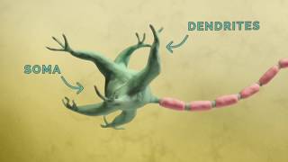
Now, using a suite of novel tools Janelia researchers have pinpointed the group of neurons in the nerve cord – a structure analogous to our spinal cord – that produce and pattern the fly’s two major courtship songs. They’ve also measured neuronal activity in these cells while flies were singing to understand how these neurons control each type of song.
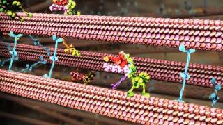
The first video shows an example trial of calcium imaging of dPR1 neurons. Top left: a bottom view of the fly. The left side of the image corresponds to the right side of the fly. Bottom left: the microphone signal for the right wing. Top right: raw fluorescent signals averaged across z-planes. The left side of the image corresponds to the left side of the ventral nerve cord. Bottom right: schematic of the imaged volume. Red boxes represent the timing of optogenetic stimulation. Optogenetic stimulation acutely increased calcium signals of dPR1 neurons as well as the amount of song, consistent with a role of dPR1 in song production.
The second video shows an example trial of calcium imaging of TN1A neurons. Top left: a bottom view of the fly. The left side of the image corresponds to the right side of the fly. Bottom left: the microphone signal for the right wing. Top right: raw fluorescent signals averaged across z-planes. The left side of the image corresponds to the left side of the ventral nerve cord. Bottom right: schematic of the imaged volume. Red boxes represent the timing of optogenetic stimulation. Calcium signals of individual TN1A neurons increased in response to optogenetic stimulation of the song driver.
Credit: Shiozaki et al. DOI: 10.1101/2022.12.14.520499