Published On Feb 3, 2014
Anatomy of Heart Attacks. This video is available for instant download licensing here: https://www.alilamedicalmedia.com/-/g...
©Alila Medical Media. All rights reserved.
Support us on Patreon and get FREE downloads and other great rewards: patreon.com/AlilaMedicalMedia
All images/videos by Alila Medical Media are for information purposes ONLY and are NOT intended to replace professional medical advice, diagnosis or treatment. Always seek the advice of a qualified healthcare provider with any questions you may have regarding a medical condition.
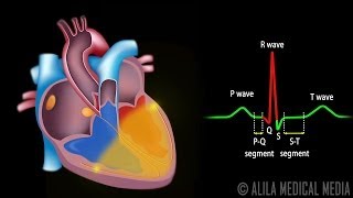
Myocardial infarction, commonly referred to as heart attack, is the sudden death of part of the heart muscle due to loss of blood flow. This occurs when one of the coronary arteries -- the arteries that supply blood to the heart -- is blocked. The blockage is commonly due to atherosclerosis - cholesterol plaques/fat deposits on the wall of blood vessels. As the plaque builds up, the vessel becomes narrow restricting blood flow. Under stress, the plaque may rupture. This triggers formation of blood clot on top of the plaque leading to complete blockage of blood flow. When this happens in a coronary artery, the downstream patch of the myocardium dies from lack of oxygen. Weaken heart muscle may disrupt electrical activity of the heart and subsequently cause cardiac arrest.
Coronary angioplasty is a non-surgical procedure used to open narrowed or blocked coronary arteries. It can also be performed as an emergency treatment for myocardial infarction. The first part of the procedure is to localize the site of blockage. This part is called cardiac catheterization. A guiding catheter is inserted through the femoral artery at the groin and threaded all the way to the aorta. The tip of the catheter is placed at the beginning of the coronary artery to be investigated. A radio-opaque dye is injected through the catheter into the coronary artery. This enables real-time visualization of the artery using X-ray imaging. A narrowed part of an artery would appear as a bottle neck on an x-ray image. After the location of narrowed artery is identified, angioplasty can begin. A guidewire with a deflated balloon is inserted and pushed to the location of blockage. The balloon is inflated to crush the plaque. At the end of procedure, the balloon is again deflated and removed together with all catheters and guidewire. In some cases, a stent is inserted together with the balloon, inflated and left on place of the plaque to keep the artery open permanently.